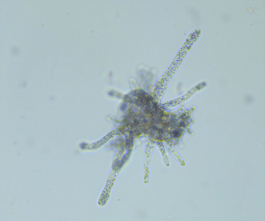
Have you ever considered how a single-celled organism can reveal the fundamental organisation of life when preserved on a slide and studied under a microscope?
In biology education, access to stable and reliable teaching materials is essential for building accurate observational skills. With increasing emphasis on microscopy-based learning, educators rely on high-quality resources such as an Amoeba proteus slide to help students study cellular organisation without variability from living conditions.
At Blades Biological, we supply prepared materials, including prepared microscope slide sets, Protozoa slide online selections, and fungi microscope slides, designed to support structured laboratory teaching with consistent results.
This article examines how preserved Amoeba proteus specimens support classroom learning, focusing on cellular structure, teaching applications, and how educators can integrate Protozoa slide material into microscopy sessions for effective instruction.
The value of preserved protozoan slides in biological teaching
In many classrooms, variability in live cultures can limit consistency during microscopy sessions. Preserved specimens address this challenge by providing stable, non-living material that retains key structural features for repeated examination.
An Amoeba proteus slide offers educators a dependable specimen that highlights internal organisation at the unicellular level. Unlike live samples, preserved material allows prolonged observation without time constraints, making it easier for students to focus on structural identification and annotation.
Key advantages of preserved microscope slide sets include:
- Long-term stability with no requirement for maintenance or feeding
- Consistent visibility of cellular structures across multiple sessions
- Safe handling within standard laboratory environments
- Reduced variability between student observations
- Compatibility with standard school and college microscopes
Within Protozoa slide collections, Amoeba proteus remains one of the most commonly used examples for demonstrating eukaryotic cell organisation due to its clearly defined internal components.
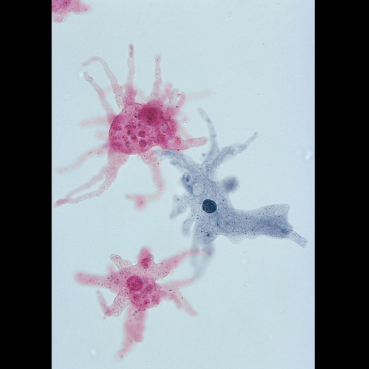
Cellular structure observable in Amoeba proteus preparations
When observing Amoeba proteus under a microscope, students are able to identify a range of internal structures that illustrate core biological principles. Although the specimen is preserved, the structural integrity remains suitable for teaching cellular organisation and compartmentalisation.
Key identifiable features include:
- Cell membrane: The outer boundary that defines cellular limits and maintains structural form
- Cytoplasm: A granular matrix that provides context for internal organisation
- Nucleus: A dense structure containing genetic material, often clearly visible in stained preparations
- Vacuole regions: Areas indicating intracellular compartmentalisation
- Cytoplasmic inclusions: Variable structures that assist in demonstrating cellular diversity
The clarity of these features makes the Amoeba Proteus slide particularly effective when introduced alongside other Protozoa slide material. Educators often compare multiple specimens within prepared microscope slide sets to highlight differences in cellular organisation among protozoans.
By using Protozoa slide online resources as part of lesson planning, instructors can ensure students are exposed to a wide range of unicellular models that reinforce theoretical knowledge from textbooks.
Practical use in laboratory sessions
Integrating preserved slides into practical lessons requires structured observation techniques. A consistent approach helps students extract maximum educational value from each specimen.
Recommended classroom workflow:
1. Microscope calibration
Students begin by adjusting illumination and focus to ensure optimal clarity. Low magnification is typically used initially.
2. Initial survey of specimen
The Amoeba proteus slide is scanned to identify general shape and distribution of cellular components.
3. Progressive magnification
Higher magnification is used to examine internal structures such as the nucleus and cytoplasmic regions.
4. Structured annotation
Students label observed structures, often using worksheets designed around Protozoa slide material.
5. Comparative analysis
Observations are compared with other prepared microscope slide sets, including fungi microscope slides, to highlight differences between unicellular and multicellular organisation.
This structured approach ensures that observing Amoeba proteus becomes a focused exercise in identification and analysis rather than a superficial viewing activity.
Supporting biological concepts through microscopy
Preserved protozoan slides play a significant role in reinforcing several core biological concepts within secondary and tertiary education.
1. Cell organisation
Students can directly observe how a single cell contains all necessary components for independent function, reinforcing the concept of unicellular life forms.
2. Eukaryotic structure
The presence of a distinct nucleus in Amoeba proteus supports teaching of eukaryotic cell classification.
3. Structural differentiation
When compared with fungi microscope slides or other Protozoa slide specimens, students can distinguish between cellular organisation patterns.
4. Microscopy skills development
Repeated use of microscope slide sets strengthens technical skills such as focusing, magnification control, and specimen interpretation.
These learning outcomes are strengthened by the consistency of preserved specimens, which eliminate environmental variation and allow educators to focus on core scientific principles.
Enhancing curriculum delivery with prepared slide collections
Prepared teaching materials simplify lesson planning and improve consistency across different student groups. A well-organised set of microscope slide sets allows educators to deliver structured sessions without the unpredictability of live culture preparation.
Within Protozoa slide online catalogues, Amoeba proteus remains a central specimen due to its clarity and educational value. When integrated with fungi microscope slides and other biological materials, it supports a broader curriculum covering cell biology, taxonomy, and microscopy techniques.
Educators benefit from:
- Reduced preparation time before laboratory sessions
- Standardised specimens across multiple teaching groups
- Reliable visibility of cellular structures
- Alignment with examination board requirements
This consistency is particularly valuable in institutions where multiple classes require identical learning experiences.
What can a preserved cell reveal about life at its most fundamental level?
When carefully prepared and observed, even a non-living specimen can provide detailed insight into biological organisation and cellular structure.
The Amoeba proteus slide remains one of the most effective teaching tools for demonstrating unicellular architecture. Its preserved form allows repeated observation without degradation, making it ideal for structured classroom analysis. When used alongside other Protozoa slide materials and prepared microscope slide sets, it strengthens student understanding of comparative cell biology and microscopic techniques.
At Blades Biological, we provide a wide range of educational microscopy resources, including Protozoa slide collections, fungi microscope slides, and carefully prepared teaching sets designed for consistent classroom use. As one of the leading suppliers of high-quality preserved specimens, including Carolina Biological materials, we support educators in delivering reliable and structured practical sessions.
For institutions aiming to strengthen their microscopy curriculum, our selection of prepared microscope slide sets offers dependable resources for teaching cell structure and biological classification.