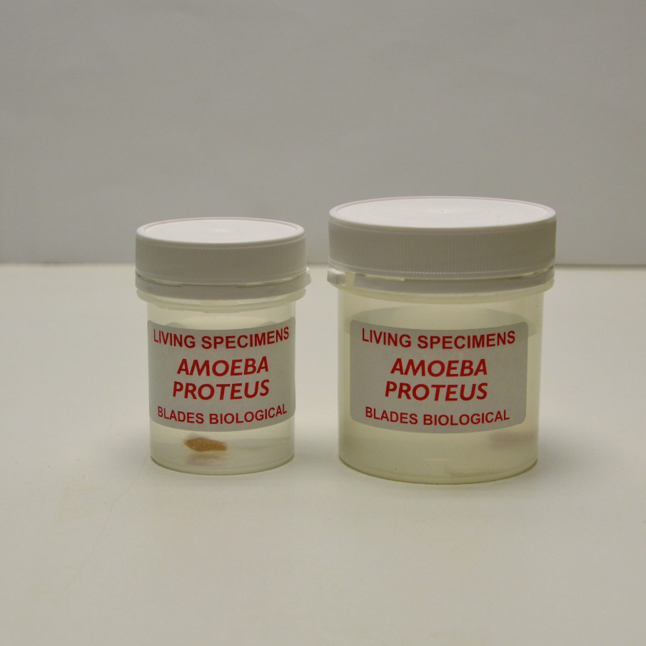
What allows students to understand cellular organisation more effectively than observing biological structures under a microscope with real, well-prepared specimens?
In many biology classrooms, a recurring limitation is the gap between theoretical diagrams and authentic microscopic observation. Textbook illustrations of protozoan organisation often fail to reflect the complexity seen in laboratory preparations.
This is where well-maintained biological resources such as Amoeba proteus slide materials and related microscopy specimens become essential in supporting structured microscopy-based learning.
Suppliers such as Blades Biological provide educators with reliable laboratory materials, including Protozoa slide online resources, prepared microscope slide sets, and fungal and algal teaching cultures that strengthen practical understanding.
This article examines how live Amoeba proteus cultures enhance laboratory teaching, how they support microscopy-based learning outcomes, and how educators can integrate Protozoa slide materials and microscope slide sets into structured practical sessions.
Strengthening Practical Biology Through Direct Microscopic Study
Practical biology teaching has long relied on observation as a foundation for conceptual understanding. When learners engage directly with prepared microscope slide sets, they are able to connect structural features with biological function in a controlled setting.
Amoeba proteus preparations are particularly valuable because they represent a single-celled eukaryotic organism with clearly defined internal structures. When used alongside Protozoa slide collections, they help learners compare cellular organisation across different protozoan groups.
Key educational advantages include:
- Reinforcement of cellular structure identification, including nucleus, cytoplasmic regions, and vacuolar components
- Improved understanding of protozoan classification within microbiology curricula
- Support for structured microscopy-based learning activities in both secondary and advanced biology programmes
- Opportunity to compare protozoa with other microorganisms from microscope slide sets
- Integration with fungi microscope slides to highlight differences between unicellular and filamentous life forms
By working with consistent, well-prepared materials, educators can maintain continuity across practical sessions and reduce variability in classroom outcomes.
Role of Amoeba proteus Specimens in Laboratory Teaching
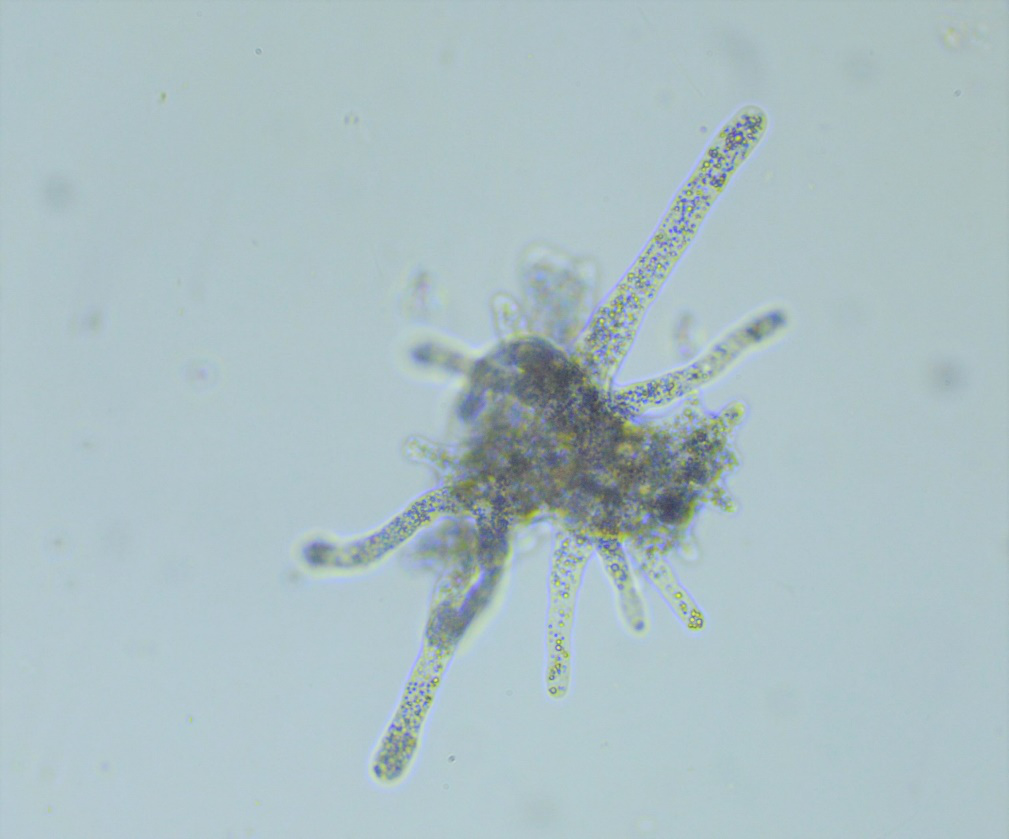
Amoeba proteus remains one of the most widely used protozoan organisms in educational microscopy due to its distinct cellular organisation. In laboratory settings, Amoeba proteus slide materials are commonly used to introduce learners to eukaryotic unicellular structure before progressing to more complex microbial systems.
In structured teaching environments, these specimens help demonstrate:
- Organisation of cytoplasm into functional zones
- Presence of a defined nucleus observable under standard magnification
- Variations in internal vacuolar structures associated with cellular processes
- Relationships between morphology and biological function
When incorporated into prepared microscope slide sets, Amoeba proteus samples provide a consistent reference point for learners to revisit during revision or comparative analysis tasks.
Educators often combine these slides with Protozoa slide sets to broaden comparative study, allowing learners to distinguish between different protozoan genera based on structural characteristics.
Integrating Prepared Microscope Slide Sets into Curriculum Design
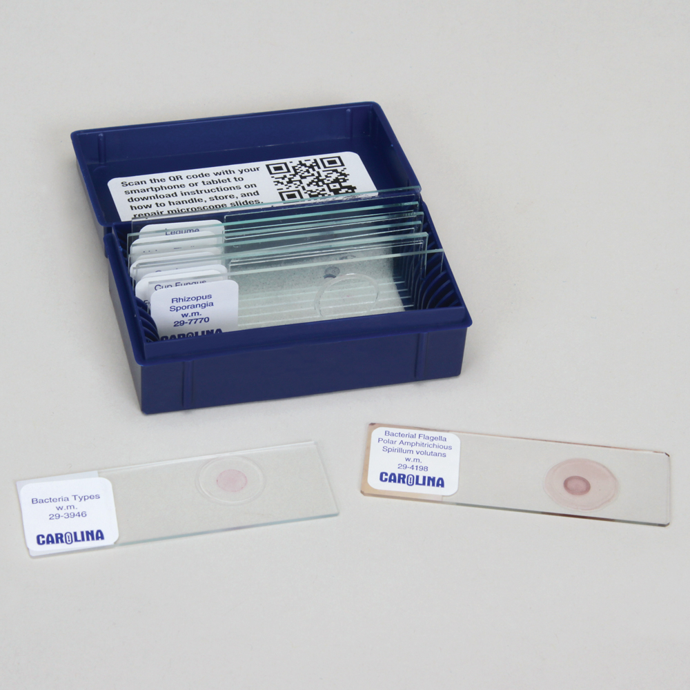
Microscopy-based learning is most effective when supported by structured and reliable teaching materials. Prepared microscope slide sets allow educators to standardise classroom observation while ensuring that each learner has access to equivalent specimen quality.
Typical uses in biology curricula include:
1. Introductory Microscopy Training
Students can develop foundational skills in focusing, magnification adjustment, and specimen interpretation using consistent slides. Protozoa slide sets are especially effective for early-stage learners.
2. Comparative Cellular Biology
Amoeba proteus preparations can be compared with other protozoan organisms, fungi microscope slides, and algal materials to highlight differences in cellular organisation.
3. Assessment-Based Observation Tasks
Teachers can design structured observation sheets where learners identify key cellular structures from microscope slide sets, reinforcing observational accuracy.
4. Extended Laboratory Projects
Advanced learners can examine multiple Protozoa slide samples to classify organisms based on structural traits and functional adaptations.
By standardising specimen quality, prepared microscope slide sets reduce variability and improve the reliability of student observations.
Enhancing Microscopy-Based Learning Outcomes
Microscopy-based learning remains one of the most effective methods for teaching biological structure at the cellular level. However, its success depends heavily on specimen clarity, consistency, and relevance to curriculum outcomes.
Amoeba proteus slide materials contribute to this by providing clear examples of unicellular organisation. When paired with fungi microscope slides and Protozoa slide online resources, educators can create a comprehensive microscopy programme that covers multiple biological kingdoms.
Key learning outcomes supported include:
- Accurate identification of cellular structures under light microscopy
- Improved understanding of unicellular organism organisation
- Development of comparative analysis skills across biological specimens
- Strengthening of laboratory documentation and observational reporting
- Reinforcement of classification principles in microbiology
These outcomes are particularly important in examination-focused curricula where practical understanding supports theoretical knowledge.
Supporting Broader Microbiological Studies in Education
Beyond protozoa, microscopy-based learning often extends into fungal and algal studies. Fungi microscope slides provide valuable contrast to protozoan samples, particularly in demonstrating filamentous structures and reproductive forms.
When used together with Amoeba proteus slide materials and other Protozoa slide sets, educators can guide learners through structured comparisons such as:
- Unicellular versus multicellular organisation
- Eukaryotic cellular diversity across biological groups
- Structural adaptation to environmental conditions
- Variation in internal cellular complexity
This comparative framework helps learners build a more coherent understanding of microbiology and cell biology as interconnected disciplines.
Practical Considerations for Laboratory Use
For effective implementation of microscopy-based learning, consistency and preparation are essential. Educators often rely on microscope slide sets and prepared microscope slide sets to ensure that classroom sessions proceed smoothly without unnecessary variability.
Best practice considerations include:
- Ensuring slides are clearly labelled for accurate identification
- Structuring lessons to progress from simple to complex organisms
- Combining Protozoa slide materials with fungal and algal specimens for breadth
- Using repeated observation tasks to reinforce recognition skills
- Maintaining organised storage for long-term usability of slide collections
These approaches help maintain a structured laboratory environment and support effective teaching delivery.
Advancing Classroom Microscopy with Structured Specimen Use
Could microscopy teaching become more effective if learners consistently worked with reliable, well-prepared biological specimens?
The answer increasingly depends on the quality and organisation of teaching resources. Amoeba proteus slide materials, Protozoa slide sets, fungi microscope slides, and broader microscope slide sets collectively provide educators with the tools needed to build structured, progressive learning experiences.
As microscopy remains central to biological education, the use of standardised, high-quality specimens continues to support deeper understanding of cellular organisation and microbial diversity.
For educators seeking dependable laboratory materials, Blades Biological offers a comprehensive range of prepared resources designed to support microscopy-based learning across multiple levels of study.