What to Expect from a Preserved Amoeba Proteus Slide in the Lab
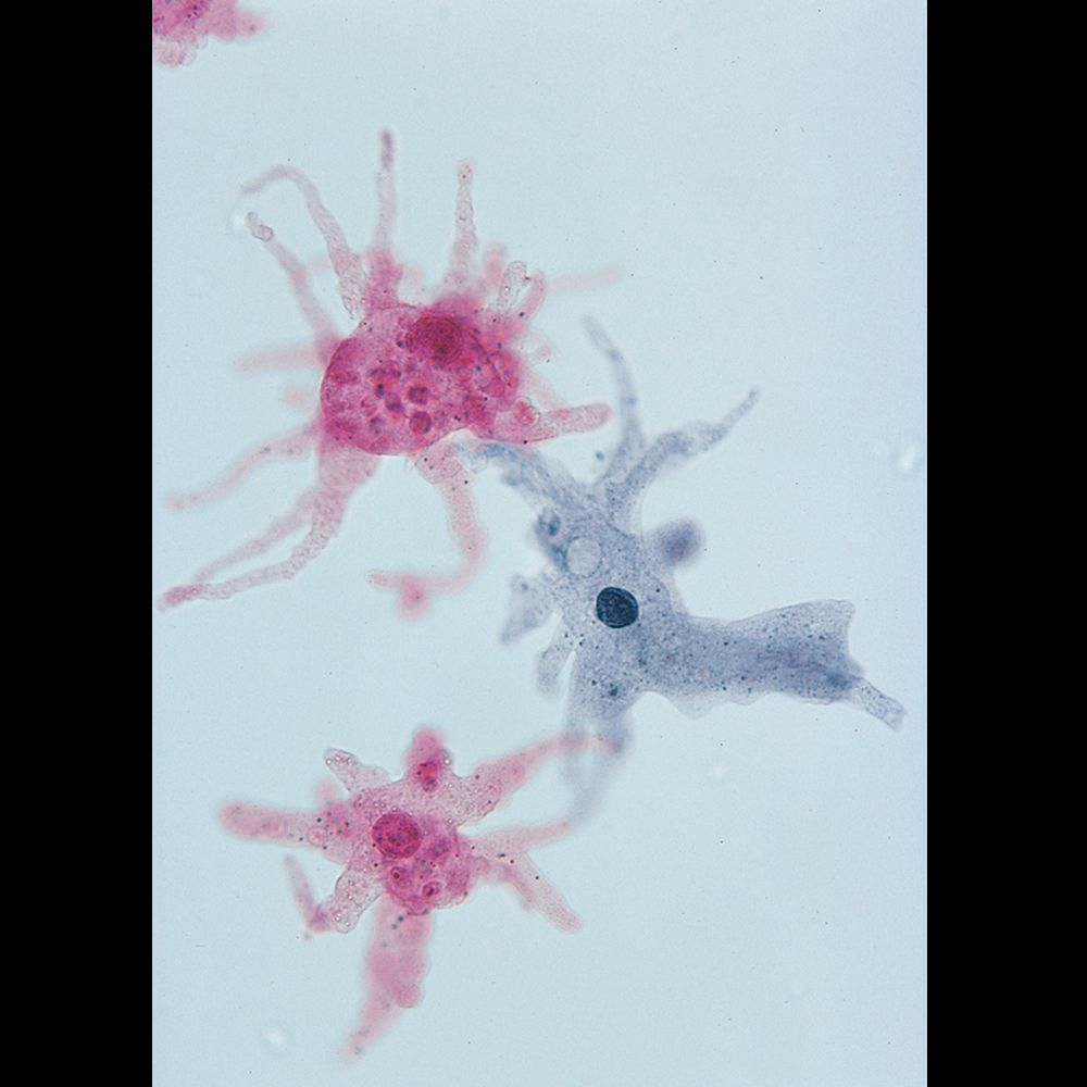
How do we teach cell biology effectively when most students have only ever seen cells in diagrams? Prepared microscope slide sets featuring preserved protozoa offer an essential solution. As schools aim to meet the microscopy components of the KS4 and post-16 science curricula, having access to high-quality preserved specimens—especially classic teaching organisms like Amoeba proteus—can transform how students understand the internal complexity of single-celled life.
At Blades Biological, we supply scientifically prepared slides that support these objectives without the variability or maintenance challenges of live cultures. As a trusted UK supplier of preserved materials, including exclusive access to Carolina Biological’s preserved specimens, we offer durable and educationally valuable protozoa slides suitable for repeated classroom use.
This blog will outline how Amoeba proteus slides are prepared, what students can expect to observe, and how these resources support microscopy education at secondary and further education levels.
Preparation of the Amoeba proteus Slide
Prepared Amoeba proteus slides are created by fixing the organism with chemical preservatives to retain cellular integrity, followed by mounting under cover glass in a stain medium. This ensures key structures remain visible and sharply defined for microscopic observation over time.
Read our insightful blog, “Standardising Microscope Slide Use in Cell Biology Practical Instruction.” (Please link to TLLA1)
What You’ll See Under Magnification
When viewed through a compound microscope, Amoeba proteus reveals several distinctive internal structures that support the teaching of cellular anatomy:
- Nucleus– often centrally located, visible as a dark, round body
- Contractile vacuole– involved in osmoregulation, appearing as a clear circular structure
- Cytoplasm– with distinguishable ectoplasm and endoplasm layers
- Pseudopodia– though fixed, their shape shows past projections used for movement and feeding
These features help bridge the gap between textbook diagrams and real biological structures.
Teaching Applications at KS4 and Above
Preserved protozoa specimens are integral to microscope slide sets that reinforce:
- Cell structure identification
- Microscopy handling and stain interpretation
- Comparisons between unicellular organisms and multicellular tissue slides (e.g. fungi microscope slides)
Using a well-prepared protozoa slide—such as Amoeba proteus—gives students hands-on context to support core learning outcomes in cell biology and practical skills in microscopy.
Ready to Improve Microscopy Teaching with Reliable Slides?
What’s the value of showing students a cell they can actually see in full structural detail? A preserved Amoeba proteus slide offers a dependable and reusable teaching tool that demonstrates core cellular structures in a real biological context. If you’re looking for microscope slide sets that align with British science curricula and support repeated classroom use, Blades Biological is one of the best sources available.
We offer a comprehensive range of prepared slides, including fungi microscope slides and Protozoa slides online, backed by over 30 years of specialist experience. Browse our prepared microscope slide sets and order today to bring accuracy and clarity to every lesson.